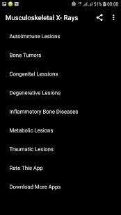
Musculoskeletal X- Rays Interpretation
x-ray interpretation guide app
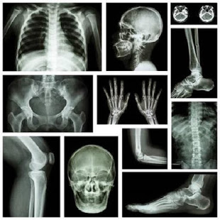
Musculoskeletal X-ray - General principles :
Systematic approach
Patient and image details
Bone and joint alignment
Joint spacing in x-ray interpretation
Cortical outline
Bone texture in chest x ray interpretation
Soft tissues
Although the system for viewing X-rays of bones and joints varies depending on the anatomy being examined, there are some broad principles which can be applied in a number of situations.A systematic approach involves checking alignment of bone structures, joint spacing, integrity of bone cortex, medullary bone texture, and for abnormalities of any visible surrounding soft tissue structures.
Patient and image details in dental x ray interpretation
Start by checking you are looking at the correct image. The patient's details should be checked and the date and time of the X-ray noted. The skeletal system is symmetrical, so it is particularly important to be sure you are looking at the correct side.
Bone and joint alignment
Loss of alignment may be due to a bone fracture or a joint dislocation. Both are associated with soft tissue injury that may not be directly visualised.
Joint spacing may be narrowed due to cartilage loss or widened due to dislocation/dissociation.
Cortical outline
Careful scrutiny of the bone cortex is required because a check that is too brief will lead to incorrect or incomplete diagnosis.In the context of trauma the clinical features of a significant injury may be masked by other injuries. Remember to be systematic, and if you spot one abnormality, do not stop until you are sure you have focused on all areas of the anatomy shown.
Bone texture
In some bones a fine matrix of fine white lines (trabeculae) is seen. Occasionally bone injury or disease will result in abnormality of this texture.
Soft tissues
Scrutinizing the soft tissues can often provide helpful information.Not uncommonly an abnormality of soft tissues is more obvious than a bone injury, or may even imply a bone injury that is not visible at all.
Confidence in assessing musculoskeletal system X-rays comes from experience and a knowledge of normal appearances. All patients are different, so being sure of the distinction between normal and abnormal is often difficult.Here are some principles that may help you to determine if a finding is normal.
2 views
In the context of trauma at least 2 views of the body part in question are usually required. If looking for specific disease entities, for example erosions in rheumatoid arthritis, this may be less important. In some cases, such as possible scaphoid injury, more than 2 images are required.2 views are better than 1
Compare with other side
Images of the asymptomatic contralateral side to a suspected abnormality are not routinely acquired for assessment of all bones or joints.If an old image of the contralateral side is available, or if the other side is included as standard (for example hip/pelvis), then comparison between symptomatic and asymptomatic appearances can be very helpful.
Compare current with previous images
The 'old X-ray' is said to be the 'cheapest test in radiology.'
If you are uncertain of an abnormality and there is an old image available of the area in question, then ALWAYS look at it. Doing this often increases diagnostic confidence, and can show progression of pathology over time.
Keep your eye on the ball
When looking at an X-ray always keep the current clinical features at the forefront of your mind.Remember - 'Treat the patient and not the X-ray!'
Look for the unexpected
Not all disease that presents with musculoskeletal symptoms is primarily related to pathology of the bones or joints. Very often pain is referred to the symptomatic area and is explained by disease of another system.For example, shoulder pain is usually due to shoulder pathology, but always keep in mind that pain may be referred to the shoulder from the cervical spine, brachial plexus or diaphragm.